

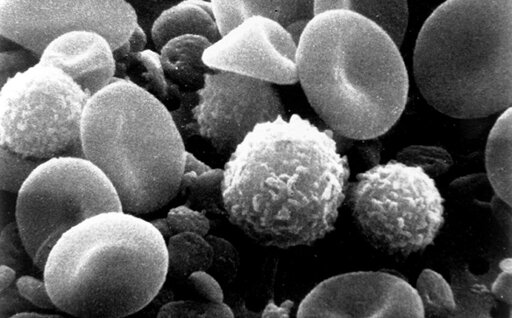
A routine blood sample contains a diverse collection of immune cells that can reveal important clues about health and disease. These cells, known as peripheral blood mononuclear cells (PBMCs), are widely used to study infections, autoimmune disorders, cancer and the immune system’s response to treatment. Scientists typically identify these cells using fluorescent labels that attach to specific surface markers. While effective, those methods provide limited information about how the cells are functioning and can alter the cells during preparation. As reported in Biophotonics Discovery, a recent study shows how advanced optical imaging can reveal a previously inaccessible layer of information: the metabolic activity of individual immune cells within a complex blood sample.
From Biology News - Evolution, Cell theory, Gene theory, Microbiology, Biotechnology via This RSS Feed.
The nondestructive nature of the technique could be especially valuable for cell therapy applications. Current methods for assessing cellular metabolism often require reagents or processing steps that can alter cell behavior. OMI, by contrast, leaves cells viable after analysis, creating opportunities to evaluate cell quality before manufacturing therapeutic products. PBMCs are commonly used as starting material for CAR T-cell therapies and other immune-cell treatments, making pretreatment assessment of cellular fitness an attractive possibility.